
Anatomy Of The Ankle Joint
When to see a doctor Summary The foot is an intricate part of the body, consisting of 26 bones, 33 joints, 107 ligaments, and 19 muscles. Scientists group the bones of the foot into the.

The Anatomy Of The Foot Anatomy Of The Foot Human Anatomy Diagram
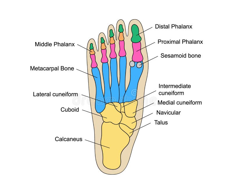
The diagram of bones in the ankle and foot is given below: Tarsal Bones The tarsal bones in the foot are located amongst tibia, metatarsal bones, and fibula. There are in all 7 bones, which fall under tarsal bones category. They are: Calcaneus or Calcaneum: To explain the term in layman's language, it is the heel bone in the skeletal system.
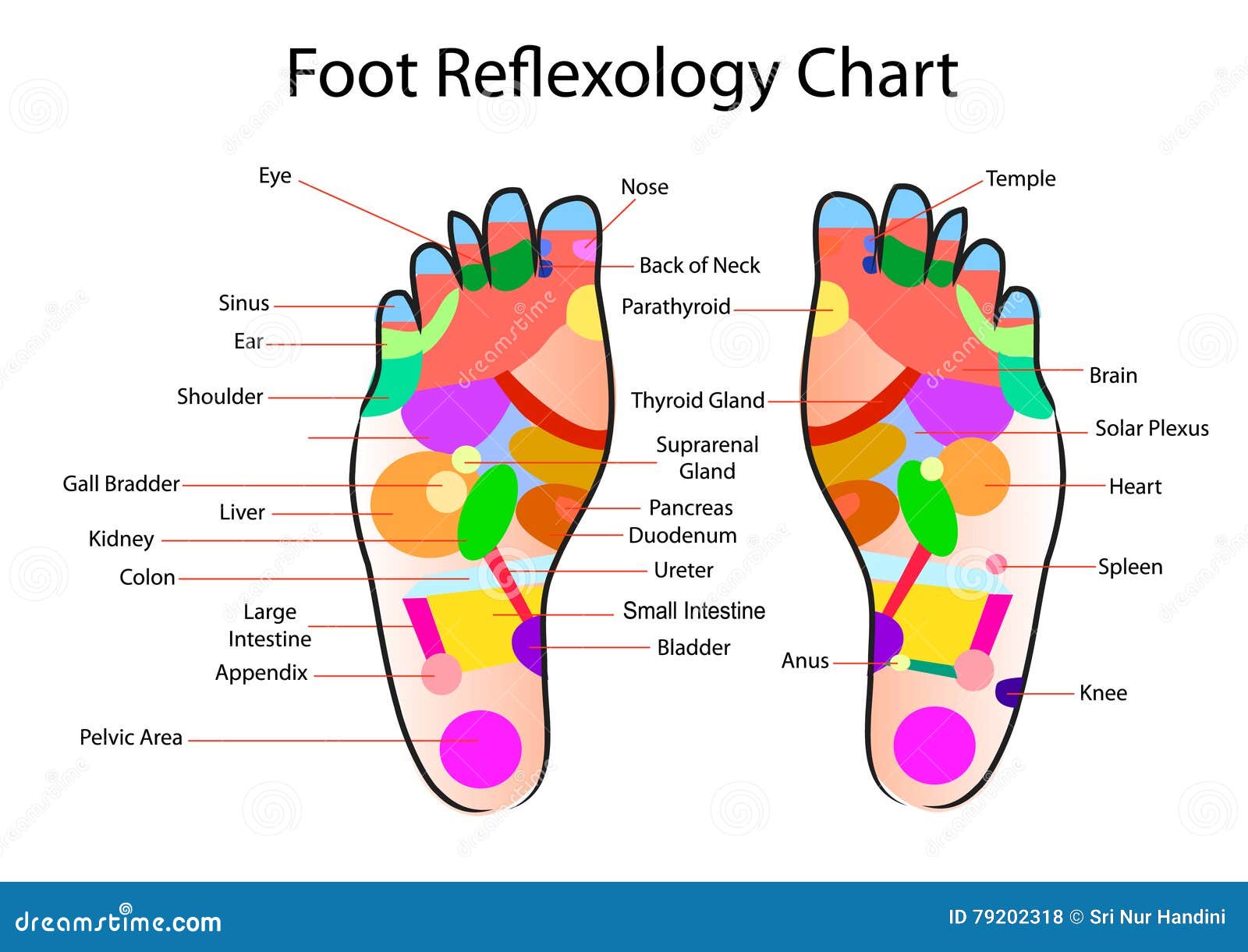
Foot reflexology chart stock vector. Illustration of accurate 79202318
In the foot, there are: 26 bones; 33 joints; more than 100 muscles, tendons, and ligaments; Bones of the foot. The bones in the foot make up nearly 25% of the total bones in the body, and they.
Foot pain relief store, foot pain diagram
The hindfoot This is comprised of the talus bone and the calcaneum. The talus connects with the tibia and fibula to form the ankle joint, and the calcaneum is the bone that forms the heel bone (ball of the foot). The calcaneum is the largest bone in the foot. The muscles, tendons and ligaments

image lateral_ankle for term side of card Ligament Tear, Ligaments And
Ankle anatomy. The ankle joint, also known as the talocrural joint, allows dorsiflexion and plantar flexion of the foot. It is made up of three joints: upper ankle joint (tibiotarsal), talocalcaneonavicular, and subtalar joints.The last two together are called the lower ankle joint. The upper ankle joint is formed by the inferior surfaces of tibia and fibula, and the superior surface of talus.

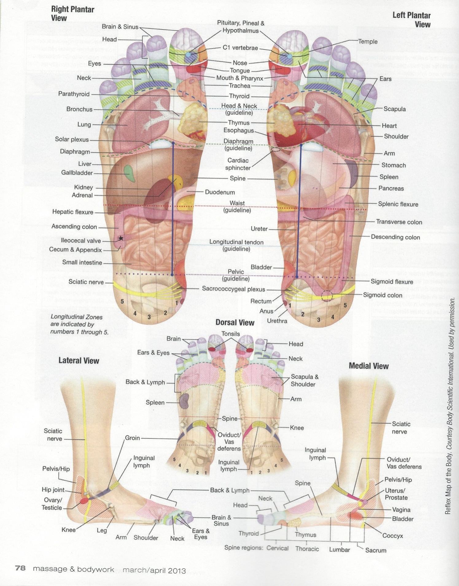
feet map of organs in body to read the organ names. It is also
The foot is the region of the body distal to the leg that is involved in weight bearing and locomotion. It consists of 28 bones, which can be divided functionally into three groups, referred to as the tarsus, metatarsus and phalanges. The foot is not only complicated in terms of the number and structure of bones, but also in terms of its joints.

Foot body organ connection. Chinese medicine. Which part of the foot
Bones of foot. The 26 bones of the foot consist of eight distinct types, including the tarsals, metatarsals, phalanges, cuneiforms, talus, navicular, and cuboid bones. The skeletal structure of.

Foot Reflexology Chart stock vector. Illustration of physical 96620033
Use these bones of the foot quizzes to master your identification skills. Overview of the bones of the foot and their divisions into the hindfoot, midfoot and forefoot. With a total of 26 bones in each foot, learning the bony anatomy of the foot is no piece of cake. That is, the memorization aspect.
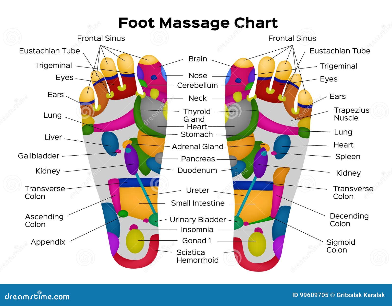
Foot Reflexology Chart with Description of the Internal Organs and Body
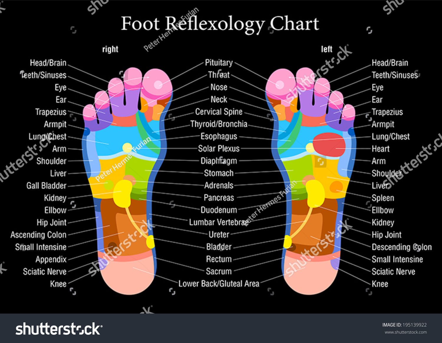
The toes and feet indicate your head and neck. Massaging your toes in foot reflexology means working your head and neck. The insides of your feet correlate to your spine. The area just underneath your toes corresponds to the chest. The thinnest part of your foot, usually found towards its center, is known as the waistline.

Foot Reflexology And Body Organs Healthy Tactic
Navicular Many of the muscles that affect larger foot movements are located in the lower leg. However, the foot itself is a web of muscles that can perform specific articulations that help.
Human Foot Bones Anatomy with Descriptions. Educational Diagram of
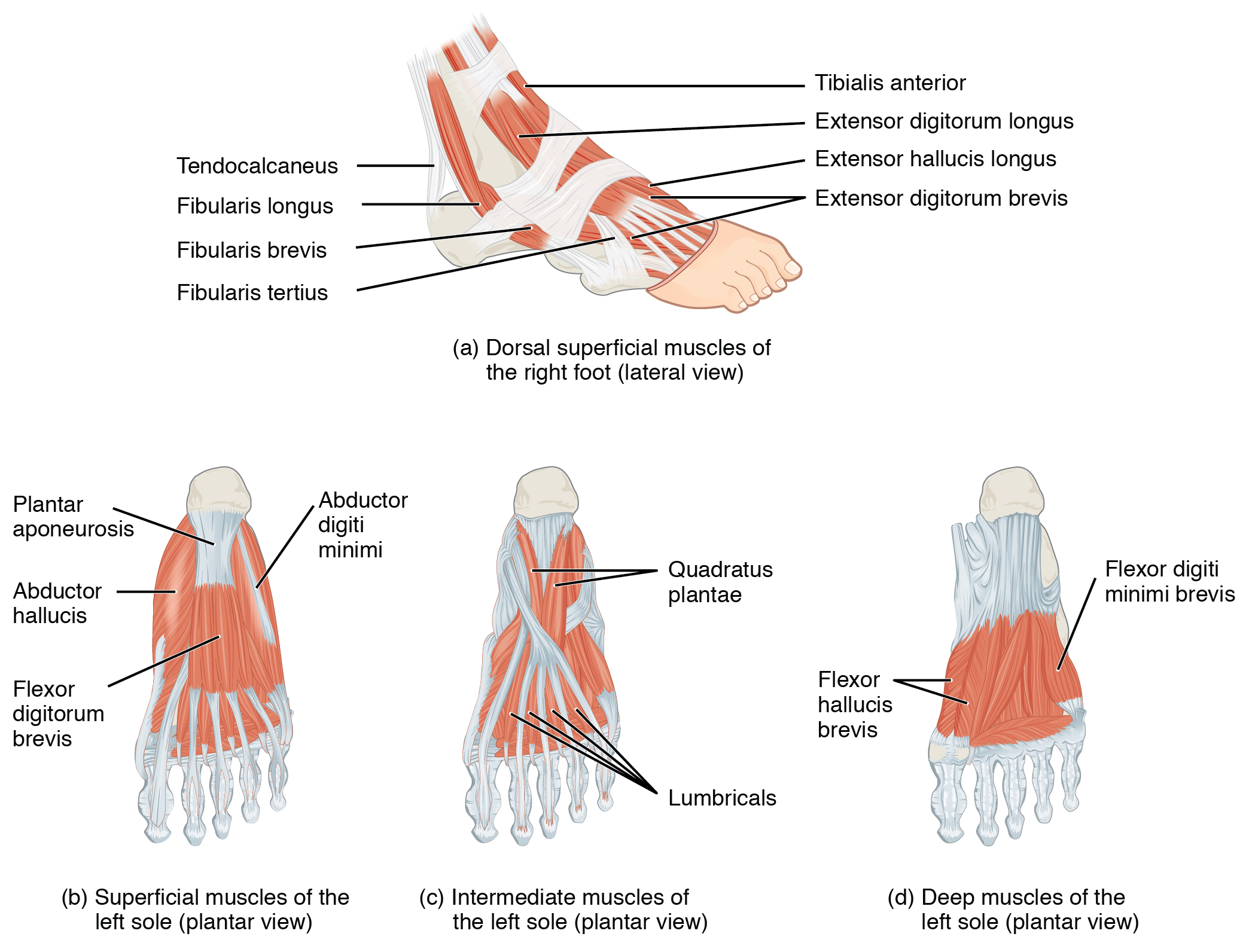
LABELED DIAGRAMS. Figure 1. Sections and Bones of the Foot A. Lateral (Left) B. Anterior (Right) Figure 2. Compartments of the Foot A. Cut Section through Mid-Foot. Figure 3. First Layer of the Foot A. Plantar View of Right Foot. Figure 4. Second Layer of the Foot A. Plantar View of Right Foot.
Foot Reflexology Chart Accurate Description Corresponding Stock Vector
Introduction A solid understanding of anatomy is essential to effectively diagnose and treat patients with foot and ankle problems. Anatomy is a road map. Most structures in the foot are fairly superficial and can be easily palpated. Anatomical structures (tendons, bones, joints, etc) tend to hurt exactly where they are injured or inflamed.
11.6 Appendicular Muscles of the Pelvic Girdle and Lower Limbs
Foot: Anatomy. The foot is the terminal portion of the lower limb, whose primary function is to bear weight and facilitate locomotion. The foot comprises 26 bones, including the tarsal bones, metatarsal bones, and phalanges. The bones of the foot form longitudinal and transverse arches and are supported by various muscles, ligaments, and.

Notes on Anatomy and Physiology Using Imagery to Relax the Weight
The foot ( pl.: feet) is an anatomical structure found in many vertebrates. It is the terminal portion of a limb which bears weight and allows locomotion. In many animals with feet, the foot is a separate [clarification needed] organ at the terminal part of the leg made up of one or more segments or bones, generally including claws and/or nails.

Human Foot Anatomy Photograph by Pixologicstudio/science Photo Library
Metatarsalgia. Metatarsalgia is a condition that causes pain in the ball of the foot. It occurs when the metatarsals, the bones in the foot, become irritated. Metatarsalgia can be caused by overuse, tight muscles, or high heels. Treatment of metatarsalgia often includes icing, stretching, and physical therapy.

Anatomy Bones Learning Skeleton Feet Chapter 5 Appendicular Skeleton
The foot is a complex part of the body that is made up of many bones, joints, muscles, ligaments, and tendons. It can easily be injured, develop diseases, or get infections. Bunions, claw toes, flat feet, hammertoes, heel spurs, mallet toes, metatarsalgia, Morton's neuroma, and plantar fasciitis are a few examples of foot problems.